Understanding the Underbite: Comprehensive Insights into Dental Misalignments, Modern Treatment Pathways, and Lifelong Oral Health Implications


An underbite—medically classified as a form of Class III malocclusion—affects an estimated 5% to 10% of the global population, making it one of the more prevalent yet frequently misunderstood orthodontic anomalies. Characterized by a dental and skeletal relationship where the lower jaw and teeth project forward beyond the upper jaw and teeth, this condition extends far beyond simple aesthetics. While society often views dental alignment through the lens of a cosmetic smile, orthodontists and maxillofacial surgeons emphasize that an untreated underbite can significantly compromise oral functionality, chewing efficiency, speech clarity, and long-term temporomandibular joint (TMJ) health.
As modern orthodontic technology advances, clinical approaches to correcting underbites have evolved significantly. From early interceptive pediatric appliances that harness active jaw growth to complex adult orthognathic surgeries paired with precision clear aligners or fixed braces, medical professionals now have a robust toolkit to address malocclusions at any stage of life. This comprehensive report examines the underlying mechanisms of underbites, the diagnostic criteria utilized by dental professionals, developmental contributing factors, historical context of orthodontic interventions, and the distinct treatment pathways available for growing children versus mature adults.
Defining the Underbite: Anatomy and Clinical Classification
To comprehend the mechanics of an underbite, one must first examine the anatomy of a standard, healthy occlusion. In a typical bite, the upper dental arch is slightly wider and positioned forward relative to the lower arch. When the jaws close, the cusps of the upper molars interdigitate with the grooves of the lower molars, and the upper front teeth (incisors) comfortably overlap the lower front teeth by a few millimeters, shielding them and maintaining facial harmony.
In an individual with an underbite, this relationship is inverted. The lower anterior teeth sit anterior to the upper anterior teeth, creating a crossbite or edge-to-edge bite relationship along the front of the mouth. Orthodontists divide these malocclusions into two primary categories: dental underbites and skeletal underbites.
A dental underbite is restricted primarily to the positioning of the teeth. In these instances, the underlying jawbones may be properly aligned relative to the skull, but individual teeth—often due to premature loss of baby teeth, delayed eruption of permanent teeth, or crowding—erupted or drifted into an abnormal forward posture.
Conversely, a skeletal underbite involves a structural discrepancy in the jawbones themselves. This occurs either when the lower jaw (mandible) outgrows the upper jaw (maxilla), when the upper jaw is underdeveloped and restricted in its forward growth, or through a combination of both skeletal factors. Determining whether an underbite is dental, skeletal, or a hybrid of both is the single most critical step in establishing an effective, long-term treatment plan.
Clinical Signs, Symptoms, and Broader Health Implications
While some underbites are immediately apparent due to a prominent lower jaw or a pronounced chin, others are subtle enough to escape casual notice, requiring professional dental imaging for accurate detection. Common clinical indicators of an underbite extend well beyond visual appearance and frequently include:
- Rapid or uneven tooth wear, particularly on the edges of the front teeth where abnormal friction occurs.
- Chronic jaw pain, soreness, or fatigue in the masticatory muscles responsible for chewing.
- Temporomandibular joint (TMJ) disorders, which can manifest as clicking, popping, locking of the jaw, or radiating facial discomfort.
- Difficulty biting off food efficiently or chewing tougher textures, which can subsequently impact nutritional intake and digestion.
- Speech impediments, such as a lisp or difficulty pronouncing specific consonants (e.g., s, z, th) due to altered tongue placement against the teeth.
- Chronic mouth breathing, often linked to structural airway restrictions associated with maxillary underdevelopment.
Left uncorrected, the physical toll of a severe underbite accumulates over time. Uneven force distribution accelerates enamel erosion, exposes dentin, and increases the risk of tooth fracture or premature tooth loss. Furthermore, chronic strain on the temporomandibular joints can lead to degenerative joint disease, chronic headaches, neck tension, and persistent facial pain that severely diminishes a patient’s overall quality of life.
Developmental Factors and Etiology
The origins of an underbite are multifactorial, stemming from a complex interplay of genetic inheritance, environmental influences, and developmental habits during early childhood.
Genetics play a dominant role in skeletal underbites. Familial traits often dictate jaw proportions and growth trajectories; individuals with a family history of prominent lower jaws or Class III malocclusions face a substantially elevated statistical likelihood of developing similar structural patterns. Anthropological studies indicate that jaw size and skeletal morphology are heavily heritable traits passed down through generations.
Beyond genetics, certain childhood habits and environmental factors can influence or exacerbate how the teeth and jaws develop. Prolonged thumb-sucking, extended pacifier use well past toddlerhood, and chronic tongue-thrusting can exert abnormal, continuous pressure on the dental arches. For instance, if a child persistently thrusts their tongue forward against the lower front teeth during swallowing or speech, it can gradually push those teeth outward. Similarly, mouth breathing resulting from enlarged tonsils or adenoids can alter tongue posture and upper jaw development, narrowing the maxilla and creating a crossbite environment that encourages a forward mandibular shift.
Diagnostic Protocols in Modern Orthodontics
Accurate diagnosis forms the bedrock of successful orthodontic intervention. When a patient presents with an underbite, a qualified orthodontist evaluates the complete craniofacial complex, rather than merely inspecting the visible alignment of the teeth.
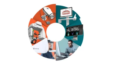
The diagnostic workflow typically encompasses a comprehensive clinical examination supplemented by advanced imaging technologies. Panoramic and cephalometric X-rays provide a two-dimensional roadmap of the skull, jawbone angles, tooth roots, and underlying skeletal structures. In modern clinical settings, three-dimensional cone-beam computed tomography (CBCT) scans and digital intraoral scans are frequently employed to generate precise virtual models of the patient’s dental arches.
Through these diagnostic tools, the orthodontist measures skeletal discrepancies, analyzes facial symmetry, evaluates airway dimensions, and predicts future growth patterns in pediatric patients. This meticulous data collection allows the clinical team to classify the malocclusion accurately and formulate a customized biomechanical strategy tailored precisely to the patient’s anatomical needs.
Chronological Evolution of Underbite Treatment Options
The management of underbites has advanced significantly over the past century, transitioning from crude mechanical wires and rigid headgear to sophisticated, computer-aided aligners and minimally invasive orthognathic surgical techniques. The appropriate treatment protocol is dictated strictly by the patient’s chronological age, skeletal maturity status, and the severity of the malocclusion.
Early Interceptive Orthodontics for Children
For decades, leading dental organizations—including the American Association of Orthodontists (AAO)—have advocated for children to undergo their first comprehensive orthodontic evaluation by age 7. At this developmental milestone, permanent incisors and first molars have typically erupted, allowing clinicians to identify developing malocclusions while the craniofacial bones are still malleable.
When an underbite is detected in a growing child, early interceptive treatment can profoundly alter the trajectory of jaw development. Clinicians frequently utilize a combination of palatal expanders and reverse-pull headgear (or facemasks). A palatal expander gently widens the upper jaw, while the reverse-pull headgear applies gentle, sustained forward pressure to the maxilla, encouraging it to grow outward to match or exceed the lower jaw.
Because children’s bones are actively growing and uncalcified at the cranial sutures, growth-guiding appliances can successfully resolve skeletal discrepancies that would otherwise require invasive surgery later in life. This proactive approach not only simplifies future phase-two orthodontic care (such as standard braces) but also establishes functional harmony during formative developmental years.
Adolescent and Adult Treatment: Braces and Clear Aligners
Once adolescent growth is complete and skeletal maturity is reached, the ability to manipulate jaw bone growth using removable appliances diminishes significantly. However, adult patients retain a wide array of highly effective treatment options designed to correct underbites through dental realignment or combined surgical-orthodontic interventions.
Fixed orthodontic appliances, commonly known as traditional braces, remain a gold standard for correcting moderate to severe underbites. Braces apply continuous, controlled biomechanical forces to reposition individual teeth within the bone. In underbite cases, braces are frequently paired with specialized intermaxillary elastics (rubber bands) attached from the upper to the lower arch, working to guide the bite into a more functional, interdigitated relationship.
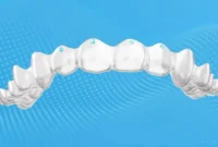
For mild to moderate underbites where the primary issue is dental rather than skeletal, clear aligner therapy has emerged as a popular and discreet alternative. Systems utilizing series of custom-molded, removable plastic trays move teeth incrementally toward their target positions. While early generations of aligners struggled with complex bite corrections, modern thermoplastic materials, precision attachment placements, and integrated power ridges allow skilled orthodontists to successfully resolve many non-skeletal underbites without traditional metal brackets.
Orthognathic Surgery for Severe Adult Skeletal Discrepancies
When an adult patient presents with a severe skeletal underbite—where the discrepancy between the upper and lower jaws is too extreme to be compensated for by tooth movement alone—orthodontic camouflage (moving teeth into abnormal angles to hide the jaw mismatch) is often insufficient or clinically contraindicated. In these instances, orthognathic surgery (jaw surgery) performed by an oral and maxillofacial surgeon is required.
The chronology of surgical underbite correction typically follows a strict, multi-stage protocol:
- Pre-Surgical Orthodontics: The patient wears braces or aligners for 12 to 18 months to align individual teeth within their respective dental arches, even if this temporarily makes the underbite appear slightly worse as the teeth are placed into their correct upright positions over their basal bone.
- The Surgical Procedure: Performed under general anesthesia in a hospital setting, the surgeon surgically cuts and repositions the upper jaw (maxillary osteotomy), the lower jaw (mandibular setback osteotomy), or both, securing them with titanium plates and screws to achieve a balanced facial profile and functional bite.
- Post-Surgical Orthodontics: Following a healing period of several weeks, the orthodontist fine-tunes the occlusion with light elastics or minor adjustments to ensure stable, long-term interdigitation.
While the prospect of jaw surgery can feel daunting to patients, modern surgical techniques, advanced three-dimensional virtual surgical planning, and improved anesthesia protocols have made the procedure safer, more predictable, and significantly more comfortable than in decades past.
Socioeconomic Implications and Public Health Perspectives
The broader implications of untreated malocclusions extend into socioeconomic and psychological domains. Studies in dental public health frequently highlight the psychological burden associated with visible dental irregularities, particularly among adolescents and young adults. Facial aesthetics and smile confidence profoundly influence social interactions, peer acceptance, and professional self-esteem.
Furthermore, the economic implications of delaying care are substantial. Interceptive treatment in childhood—though representing an upfront medical investment—frequently averts the need for expensive, high-risk surgical interventions in adulthood. From a public health standpoint, expanding public awareness regarding early screening by age 7 reduces long-term healthcare expenditure related to chronic TMJ dysfunction, restorative dental work for fractured teeth, and complex prosthodontic rehabilitation.
Navigating Treatment and Securing Professional Consultation
Because every individual’s dental anatomy, skeletal framework, and genetic predisposition are entirely unique, generalized advice cannot substitute for a clinical evaluation. Attempting to self-diagnose or postponing professional care can allow progressive malocclusions to worsen, complicating future interventions.
Dental professionals and specialized organizations emphasize that it is never too late to seek guidance. Whether evaluating a young child exhibiting early signs of a crossbite or an adult seeking to resolve a lifelong functional impairment, modern orthodontics provides tailored solutions for every stage of life.
Individuals concerned about an underbite are strongly encouraged to schedule a consultation with a qualified, board-certified specialist—such as a member of the American Association of Orthodontists (AAO). Through comprehensive diagnostic imaging, digital modeling, and expert clinical analysis, an orthodontic specialist can map out a precise, personalized treatment trajectory designed to restore oral health, optimize functional efficiency, and secure a confident, lifelong smile.