Understanding and Treating Underbites: Comprehensive Clinical Insights, Developmental Factors, and Modern Therapeutic Interventions


An underbite—medically classified as a Class III malocclusion—is a prevalent dental and skeletal condition affecting an estimated 5% to 10% of the global population. While many individuals perceive an underbite as a purely cosmetic variation in facial aesthetics, dental professionals and clinical researchers emphasize that an untreated underbite can significantly compromise long-term oral health, functional comfort, and overall quality of life. The condition occurs when the lower teeth and jaw project forward, sitting ahead of the upper teeth and maxilla when the mouth is closed in a resting or biting position.
Beyond the visible appearance of a prominent lower jaw, malocclusions of this nature can lead to uneven tooth wear, chronic temporomandibular joint (TMJ) disorders, speech impediments, and difficulties with efficient mastication and digestion. Because the etiology of an underbite can stem from dental misalignment, skeletal jaw discrepancies, or a combination of both, accurate clinical diagnosis is paramount. Modern orthodontics offers a diverse array of therapeutic interventions tailored to the patient’s age, anatomical development, and the severity of the malocclusion, ranging from non-invasive early childhood growth modification to adult orthognathic surgery.
Clinical Definition and Pathophysiology of Class III Malocclusions
To understand an underbite, it is necessary to examine the biomechanics of a healthy dental occlusion. In a standard, neutral bite—known medically as a Class I occlusion—the upper front teeth overlap the lower front teeth by a small, measured margin, and the mesiobuccal cusp of the upper permanent first molar occludes with the buccal groove of the lower permanent first molar. Furthermore, the upper dental arch is typically wider and positioned slightly anterior to the lower arch, ensuring efficient shearing and grinding of food during mastication while protecting the soft tissues of the cheeks and lips.
In contrast, an underbite represents a Class III malocclusion, where the relationship is inverted. The lower anterior teeth overlap or project anteriorly past the upper front teeth. Pathophysiologically, this misalignment can originate from two primary sources: dental factors, where the teeth themselves are tipped or angled abnormally within otherwise normally proportioned jaws, or skeletal factors, where the underlying basal bone of the mandible (lower jaw) has outgrown the maxilla (upper jaw), or conversely, where the maxilla is underdeveloped or retrognathic.
The severity of a Class III malocclusion exists on a broad spectrum. Mild cases may involve only a few lower front teeth resting edge-to-edge against the upper teeth, a condition known as an edge-to-edge bite. Moderate to severe cases feature a pronounced horizontal overjet (technically a negative overjet, where the distance between the upper and lower incisors is measured negatively) accompanied by a visible forward protrusion of the lower jaw, facial asymmetry, and compromised occlusal function.
Common Signs, Symptoms, and Secondary Health Complications
While some underbites are immediately apparent during a casual visual inspection or smiling, others are subtle, manifesting only through functional discomfort or during a comprehensive dental examination. Recognizing the clinical signs of an underbite early can prevent the exacerbation of associated health complications. Common indicators include:
- A Pronounced Lower Profile: Visually, the lower lip and chin appear to jut outward, altering the natural facial profile and symmetry.
- Difficulty with Mastication: Patients often struggle to bite into foods, tear items with front teeth, or grind food efficiently due to improper cuspid interdigitation.
- Speech Impediments: The altered relationship between the tongue, teeth, and lips can cause difficulties in articulating specific phonemes, leading to lisps or slurred speech patterns.
- Temporomandibular Joint (TMJ) Pain: Chronic stress on the jaw joints can cause clicking, popping, locking, facial myalgia, and tension headaches.
- Abnormal Tooth Wear: Because the teeth do not meet in harmony, excessive friction leads to premature enamel erosion, chipping, and sensitivity.
- Periodontal Complications: Misaligned teeth are frequently harder to clean effectively, increasing the risk of plaque accumulation, gingivitis, and localized periodontal disease.
Left unaddressed, the continuous abnormal mechanical loading on the teeth and supporting alveolar bone can lead to long-term structural damage, eventual tooth loss, and worsening skeletal asymmetries as the patient ages.
Etiological Factors: Genetics, Development, and Environmental Influences
The development of a Class III malocclusion is rarely attributed to a single cause; rather, it is typically the result of a complex interplay between genetic predispositions, hereditary skeletal traits, and environmental or functional habits during early childhood.
Genetics plays a dominant role in skeletal underbites. Familial patterns of jaw growth—such as mandibular prognathism (an excessively large lower jaw) or maxillary retrognathism (an underdeveloped upper jaw)—are frequently traceable across generations. Anthropological studies have noted specific population groups with higher hereditary propensities for Class III malocclusions, indicating a strong polygenic inheritance model governing craniofacial growth.
In addition to hereditary factors, environmental influences and childhood habits can significantly influence jaw and tooth development. Prolonged non-nutritive sucking habits, such as extensive pacifier use or thumb-sucking past the age of typical toddler development, can alter the pressures exerted on the developing dental arches. Furthermore, chronic respiratory issues leading to mouth breathing—such as enlarged tonsils, adenoids, or chronic allergies—can force the mandible to posture forward to maintain an open airway, gradually encouraging a Class III growth trajectory during formative years.
Diagnostic Protocols in Modern Orthodontics
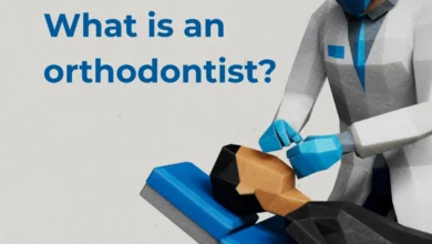
Accurate diagnosis is the cornerstone of effective orthodontic treatment planning. When a patient presents with a suspected underbite, an orthodontist conducts a thorough, multi-faceted evaluation that looks beyond the visible teeth to examine the entire craniofacial complex. The diagnostic workflow typically incorporates:
- Clinical Examination: The clinician evaluates dental relationships, facial symmetry, profile proportions, range of mandibular movement, and the health of the periodontium and temporomandibular joints.
- Digital Impressions and Study Models: Physical or digital 3D models of the dental arches allow the orthodontist to analyze the occlusion from multiple angles, measure arch length discrepancies, and calculate tooth-size ratios.
- Radiographic Imaging: Panoramic x-rays provide a comprehensive view of all teeth, roots, and surrounding bone structures. Lateral cephalometric radiographs—side-profile head x-rays—are particularly critical, as they allow clinicians to measure skeletal angles, evaluate the relative positions of the maxilla and mandible, and assess craniofacial growth patterns.
- Photographic Records: Intraoral and extraoral photographs document the baseline condition, aiding in treatment simulation and monitoring progress over time.
By synthesizing these diagnostic data points, the orthodontist classifies the underbite as primarily dental, skeletal, or a combination of both. This critical distinction dictates the appropriate therapeutic pathway.
Comprehensive Underbite Treatment Options
The therapeutic approach to correcting an underbite depends heavily on the patient’s chronological and skeletal age, the rate of remaining facial growth, and the structural severity of the malocclusion. Modern orthodontics utilizes several distinct modalities to achieve functional and aesthetic harmony.
Traditional Braces
Fixed appliances, commonly known as braces, remain a gold-standard treatment when an underbite is primarily dental in origin or involves moderate skeletal discrepancies in compliant patients. Braces apply continuous, controlled forces via brackets bonded to the teeth and archwires threading through them. This steady pressure guides the teeth into their optimal alignment. In Class III cases, braces are frequently combined with inter-maxillary elastics (rubber bands) stretched from the upper to the lower arch in specific configurations to encourage the upper teeth to move forward and the lower teeth to move backward relative to each other.
Clear Aligners
Clear aligner therapy (such as Invisalign) has evolved significantly, offering a viable alternative for mild to moderate underbites where the corrective goals can be achieved predominantly through dental movement rather than skeletal modification. Series of custom-fabricated, removable plastic trays apply incremental pressure to shift teeth systematically. While highly valued for their aesthetics and convenience, clear aligners require strict patient compliance (wearing them 20 to 22 hours per day) and are evaluated by orthodontists on a case-by-case basis to ensure they can manage the specific vectors required for underbite correction.
Early Intervention: Palatal Expanders and Reverse-Pull Headgear
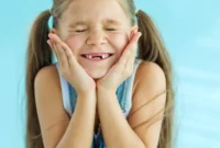
For children experiencing active craniofacial growth, early orthopedic intervention can capitalize on bone malleability to correct skeletal imbalances before they fully manifest. The American Association of Orthodontists (AAO) recommends that children undergo their initial orthodontic evaluation by age 7, as this is when permanent incisors erupt and early skeletal discrepancies become identifiable.
When a child presents with a retrognathic (underdeveloped) maxilla contributing to an underbite, clinicians frequently utilize a rapid palatal expander (RPE) combined with a reverse-pull headgear (also known as a facial mask). The palatal expander widens the upper jaw by gently separating the mid-palatal suture, while the reverse-pull headgear applies gentle, forward-directed traction to the upper jaw. Because a child’s facial bones are still fusing, this growth-modification approach can successfully stimulate forward development of the maxilla, often mitigating the need for more invasive surgical interventions later in life.
Orthognathic Surgery (Jaw Surgery)
When an underbite involves a severe skeletal discrepancy in an adult whose facial bones have finished growing, orthodontic camouflage (moving teeth to compensate for mismatched jaws) may be insufficient or functionally unstable. In these cases, a combined approach involving pre-surgical orthodontics, orthognathic surgery, and post-surgical orthodontics is indicated.
Performed by an oral and maxillofacial surgeon in coordination with an orthodontist, jaw surgery repositions the maxilla, the mandible, or both into a harmonious alignment. Pre-surgical braces align the teeth within their respective jaws independently, even if the bite looks worse temporarily. Once surgery is performed to correct the underlying skeletal bone structure, post-surgical orthodontics fine-tunes the occlusion. Although more intensive, orthognathic surgery delivers transformative results for adult patients with severe structural deformities, permanently restoring balanced facial aesthetics, airway patency, and optimal bite function.
Comparative Analysis: Pediatric Versus Adult Interventions
The physiological differences between developing pediatric patients and fully matured adult patients fundamentally shape the prognosis and methodology of underbite treatment.
| Clinical Parameter | Pediatric Patients (Under 12–14 years) | Adult Patients (Post-Growth Completion) |
|---|---|---|
| Bone Plasticity | High; bones are actively growing and malleable. | Complete; bones are dense and fully fused. |
| Treatment Modalities | Growth modification appliances, palatal expanders, functional appliances, phase-one braces. | Fixed braces, clear aligners, orthognathic surgery (if skeletal). |
| Intervention Speed | Often faster, utilizing natural growth vectors to assist correction. | Dependent on cellular remodeling rates; may take longer for skeletal adaptations. |
| Surgical Necessity | Extremely rare; growth guidance typically avoids future surgery. | Required in moderate-to-severe skeletal discrepancies where camouflage is inadequate. |
While adult treatment requires more strategic planning due to the cessation of natural craniofacial growth, advancements in dental technology ensure that adults can achieve exceptional, stable functional outcomes at any age.
Economic and Public Health Implications of Malocclusion Management
Beyond individual clinical outcomes, the management of malocclusions such as underbites carries broader public health and economic implications. Untreated severe bite discrepancies contribute to chronic healthcare utilization, including frequent consultations for temporomandibular joint dysfunction, specialized periodontal treatments for localized trauma, restorative procedures for fractured teeth, and speech therapy interventions.
Public health analyses suggest that early screening programs—such as routine orthodontic evaluations recommended by professional bodies like the AAO at age 7—reduce the incidence of complex, costly interventions in adulthood. By identifying skeletal discrepancies during the mixed dentition phase, healthcare systems and families can leverage natural growth processes, thereby lowering the lifetime economic burden associated with extensive reconstructive surgeries and chronic pain management.
Furthermore, correcting functional malocclusions improves nutritional intake by restoring efficient mastication, enhances psychosocial well-being, and eliminates speech-related barriers in professional and social environments.
Conclusion and Professional Recommendations
Underbites are well-documented, highly understood clinical conditions that, while potentially complex, respond exceptionally well to modern evidence-based orthodontic care. Because every patient’s anatomical structure, age, and developmental trajectory are unique, there is no universal template for treatment.
Healthcare professionals and dental organizations consistently underscore the necessity of personalized evaluations conducted by qualified specialists. Whether evaluating a developing child to harness natural growth vectors or exploring comprehensive adult orthodontic and surgical options to resolve a longstanding skeletal mismatch, professional oversight ensures safety, efficacy, and long-term stability.
Individuals experiencing functional discomfort, bite irregularities, or cosmetic concerns regarding their jaw alignment are strongly encouraged to consult a certified orthodontic specialist. Early diagnostic evaluation remains the most effective tool in mitigating complications, demystifying treatment options, and charting a clear, predictable course toward optimal oral health and functional harmony.